The cholera bacterium can hitchhike amoebas to avoid danger

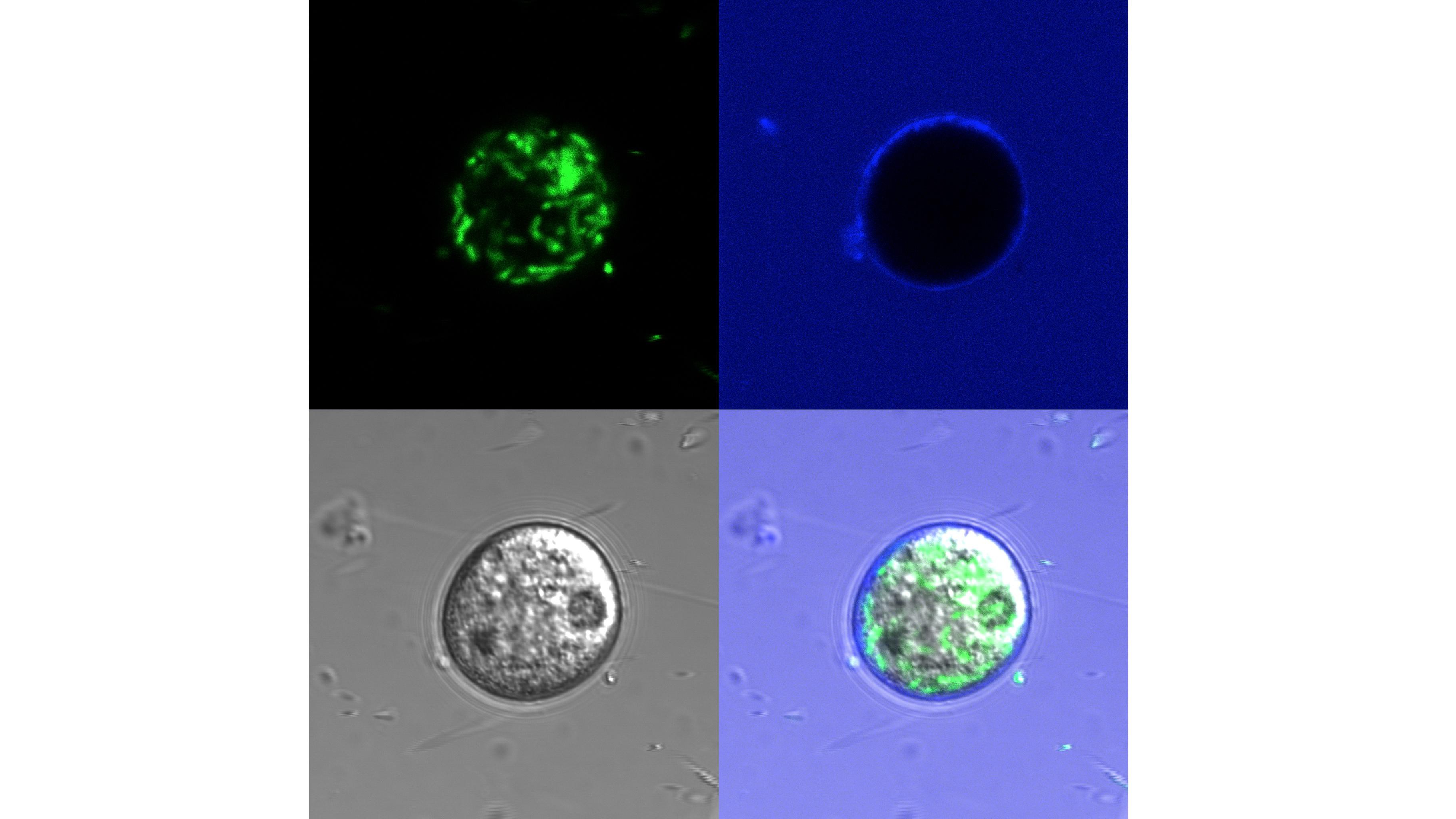
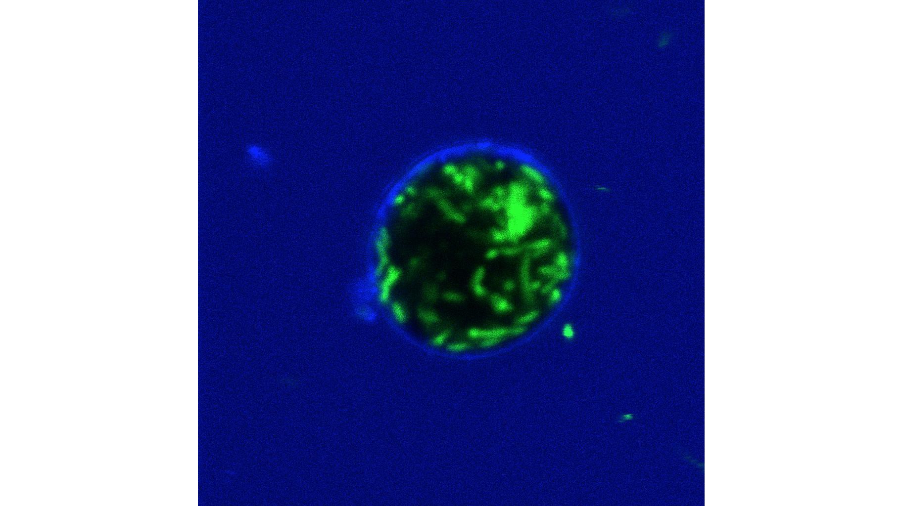
Vibrio cholerae bacteria inside the water-discharge vacuole of an amoeba © Melanie Blokesch/EPFL
EPFL scientists have discovered that the cholera bacterium can hitchhike aquatic amoebas, which might allow it to bypass threats in the surrounding water as well as the human stomach. The finding could have major implications for cholera transmission to humans.
The cholera bacterium, Vibrio cholerae, currently infects millions of people, especially in countries with poor sanitary conditions. The bacterium lives in water, e.g. oceans, ponds and rivers, where it has evolved formidable skills to ensure its survival, growth, and occasional transmission to humans. Scientists at EPFL have now uncovered a new survival strategy, wherein the bacterium “hitchhikes” a waterborne amoeba, hiding inside one of the amoeba’s organelles. V. cholerae is maintained within this compartment even when the amoeba retreats into the form of a tough cyst. In this way, the bacterium might protect itself from external stresses, including the acidic barrier of the human stomach. The work is published in the ISME Journal.
Even in its natural habitat, water, the cholera bacterium is under constant threat from other microorganisms trying to kill it. Major enemies are aquatic amoebae, which engulf the bacterium to digest it. This ongoing predation is thought to have contributed to the bacterium’s evolution. As a result of this evolutionary pressure, the bacterium has developed several virulence mechanisms that help it to adapt and survive.
Charles Van der Henst, a postdoc in Melanie Blokesch’s lab at EPFL, has now discovered an unsuspected trick that the cholera bacterium uses to survive attacks from amoebas: it hitches a ride in them. Looking at the level of single cells, the researchers studied how different strains of V. cholerae interact with the co-habiting aquatic amoeba Acanthamoeba castellanii. They found that a subset of V. cholerae can resist intracellular digestion by the amoeba. These undigested bacteria are normally released by the amoeba; but the study showed that they can also form a “replication niche” within it.
Specifically, V. cholerae establishes itself inside a compartment that the amoeba usually releases water with to regulate its osmotic pressure. The cholera bacteria colonize this water-discharge vacuole, and then grow inside it with an efficiency that remains undisturbed even when the amoeba responds to the occupation by turning into a hardened cyst (this process is known as “amoebal encystment”). Once they grow to sufficient numbers, the bacteria escape the encysted amoeba by lysing their way out of it, and killing it in the process.
"The results of the study are stunning,” says Melanie Blokesch. "This kind of intracellular localization and growth inside eukaryotic cells was not previously thought to occur for V. cholerae. The discovery certainly warrants a closer look."
The study describes a new host-pathogen interaction pathway for V. cholerae, which involves the bacterium killing its host. Since amoebal cysts are very resistant to bleach and acidic environments in general, the scientists suggest that, by hitchhiking an amoeba, V. cholerae bypasses common water disinfection strategies as well as the acidic barrier of the human stomach. Ultimately, this resistance would have a major effect on the transmission of cholera to human populations.
This work was funded by the European Research Council and the Swiss National Science Foundation.
Reference
Van der Henst C, Scrignari T, Maclachlan C, Blokesch M. An intracellular replication niche for Vibrio cholerae in the amoeba Acanthamoeba castellanii.The ISME Journal 22 September 2015. DOI: 10.1038/ismej.2015.165
Images to download