Researchers open door to stain-free labeling of cellular components
Scientists at EPFL and the Consiglio Nazionale delle Ricerche (CNR), the University Federico II, and CEINGE-Biotecnologie avanzate in Naples, Italy, have developed a new method to screen individual cells quickly and reliably without fluorescence labeling. Their work, published in the journal Nature Photonics, opens new avenues in early tumor diagnosis and drug development.
Under the microscope, healthy and unhealthy cells can be very difficult to distinguish. Scientists use stains or fluorescent tags targeting specific proteins to identify cell types, characterize their state, and study the impact of drugs and other therapies. While its impact on medicine has been transformational, the approach has its limitations. For one, tagging cells is expensive, time-consuming, and strongly dependent on the researcher’s skill. On top of that, the staining process can be detrimental to the cells under investigation.
That’s why researchers have been developing alternative ways to quickly and reliably screen individual cells. In a recent article published in the journal Nature Photonics, researchers from EPFL’s School of Engineering and colleagues from the Institute of Applied Sciences & Intelligent Systems, CNR, in Pozzuoli, Italy, present a stain-free approach capable of accurately distinguishing specific regions within living cells. Uniquely combining holographic imaging and microfluidics with neural network-based signal processing, the work paves the way for liquid biopsies for circulating tumor cell detection and high-throughput assays for drug testing.

From the phase delay to the refractive index
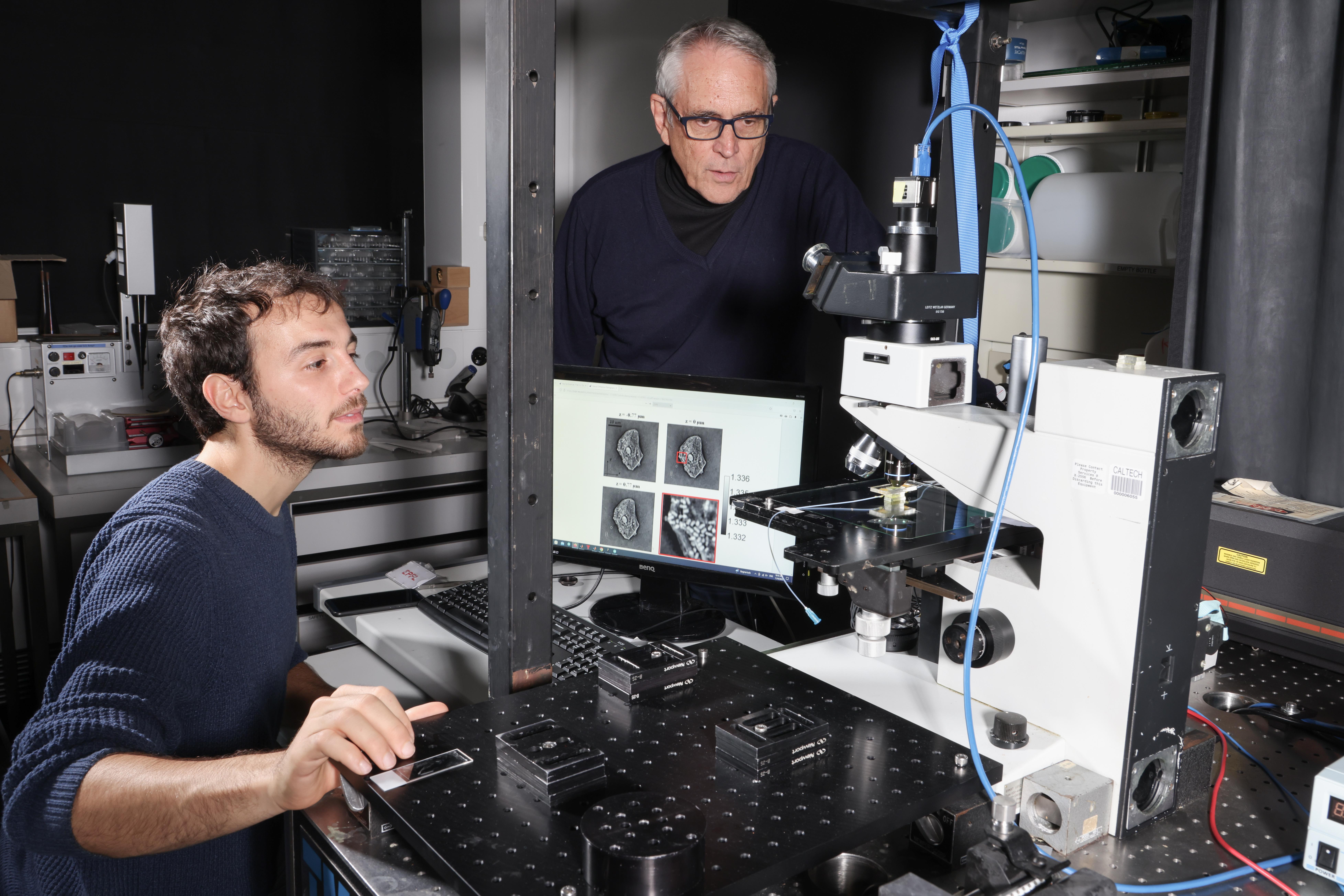
The study builds on learning tomography, a method previously developed by Demetri Psaltis and his team at the EPFL Optics Laboratory. Rather than using a microscope to create a visual image of the specimen under study, learning tomography relies on quantitative phase imaging, a holographic imaging approach that reveals the phase delay incurred as the microscope’s light beam passes through the matter that makes up the cell.
Repeating this process at several different angles and running the phase data through a neural network allowed the researchers to generate 3D maps of the refractive index of each individual voxel – each three-dimensional volume resolved by the method. “The refractive index is influenced by the density of molecules and the material,” explains Psaltis. Increasing the number of iterations further improved the accuracy of the refractive index distribution estimate.

Classifying cellular components
In their publication, Psaltis and his team present how they overcame a long-standing limitation of quantitative phase imaging approaches: the inability to identify intracellular components. “Using a self-clustering approach that groups voxels with a similar refractive index coupled with machine learning tools allowed us to assemble the clusters into shapes that we could classify. Different types of nuclei, for example, have different indices of refraction,” says Psaltis. Closing this gap paves the way for quantitative phase imaging to deliver insights previously only obtainable using fluorescence microscopy.
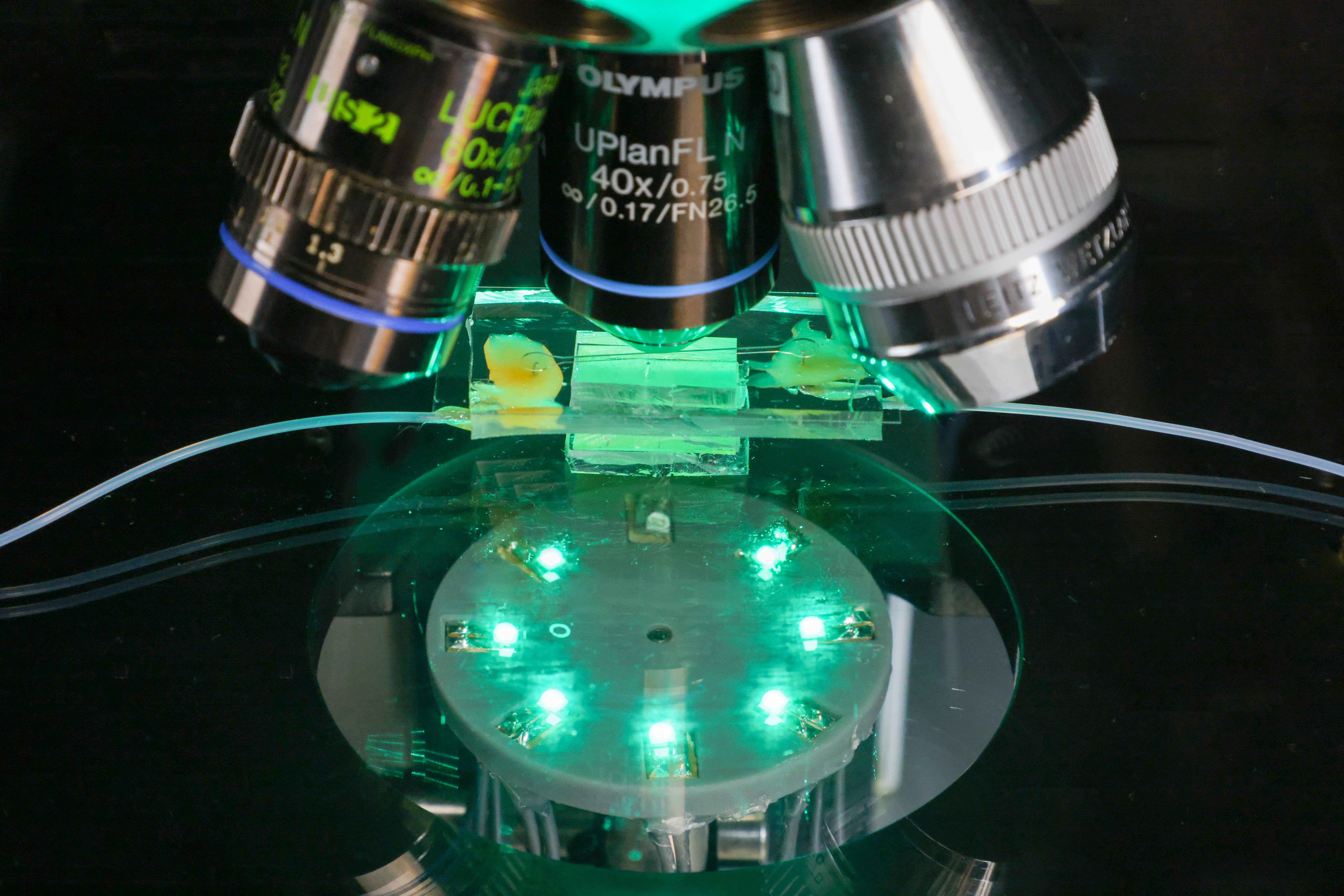
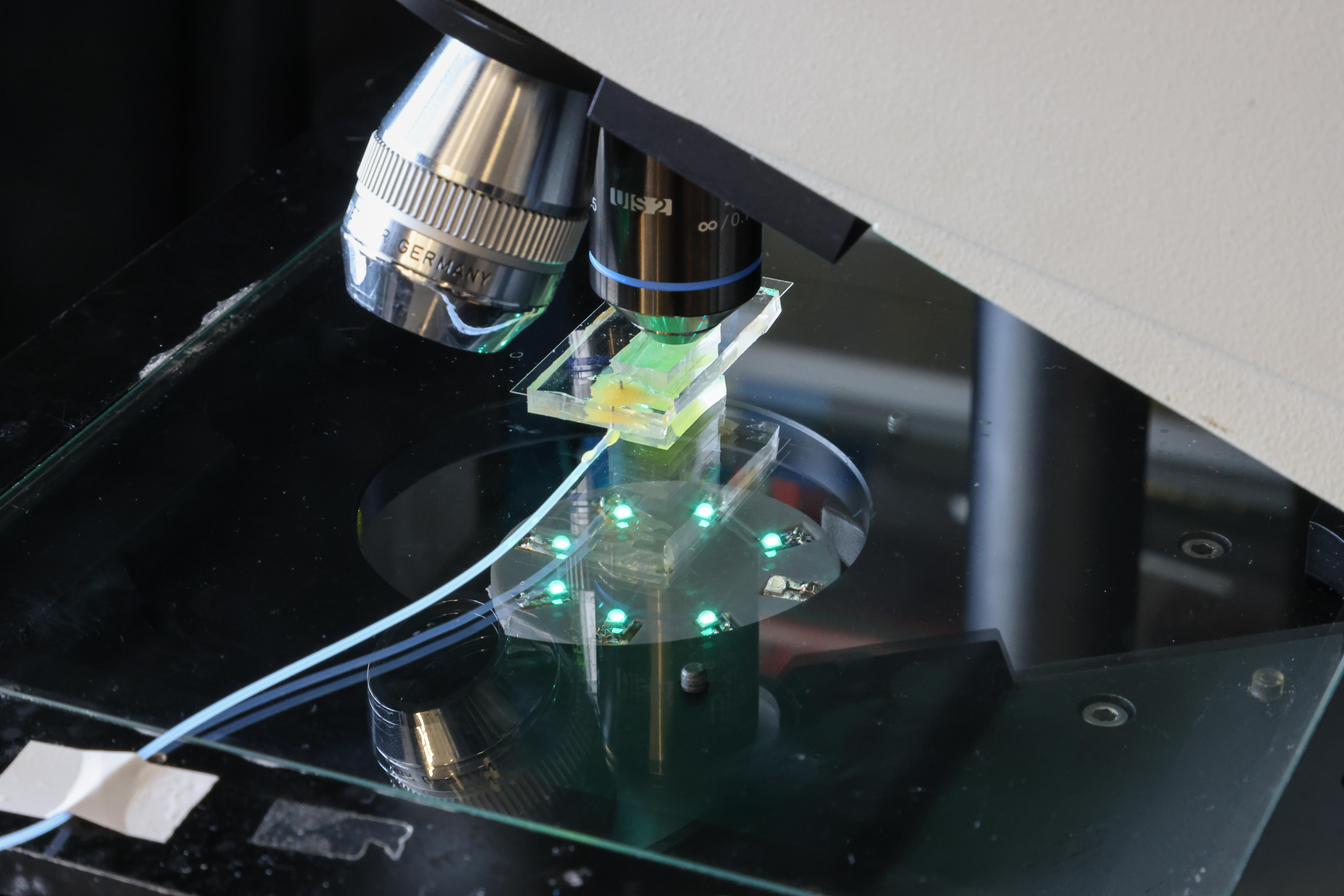
A second challenge was developing a method to screen cells that did not require immobilizing them. The solution to this challenge came from co-author Pietro Ferraro and his laboratory at CNR, who had vast experience working on in-flow tomography using lab-on-chip devices. “The idea was to put the cells in a fluidic channel 50 to 100 microns across and let the flow velocity gradient in the channel rotate the cells,” says Psaltis. “By observing the cells as they tumble along the channel using a stationary beam and detector, we can detect the phase delay, estimate the orientation of the cell, and apply our learning tomography approach to generate the 3D refractive index maps.”
“The achievable transverse resolution is of half a micron to one micron,” says Psaltis. “We can’t detect individual proteins, but we can see protein aggregates, which tend to be tens of microns across. It also let us assess the size of the nucleus and the outline of the cell, which becomes less smooth when cells become cancerous.” The researchers validated their methodology by comparing their findings with observations made using confocal fluorescence microscopy, today’s gold standard in 3D cellular imaging.

High-throughput screening of individual cells
An essential application of stain-free cell screening is liquid biopsies that allow the detection of circulating cancer cells, used both to identify cancer types in surgery and as an early diagnostic tool for cancer metastasis.
Another is drug development. Many diseases, such as Parkinson’s, are associated with cross-linked proteins. The approach developed by Psaltis and his collaborators offers a highly efficient, non-invasive way to evaluate the effectiveness of drugs designed to break down these cross-linked proteins in real-time by repeatedly running treated cells through the imaging setup. In the same way, the approach could be used to give researchers new insights into the real-time effects of pathogens on healthy cells.
According to Psaltis, future work will involve applying machine learning tools to extract biologically relevant information and concrete diagnoses from the estimated refractive index distribution.