Observing cellular activity, one molecule at a time

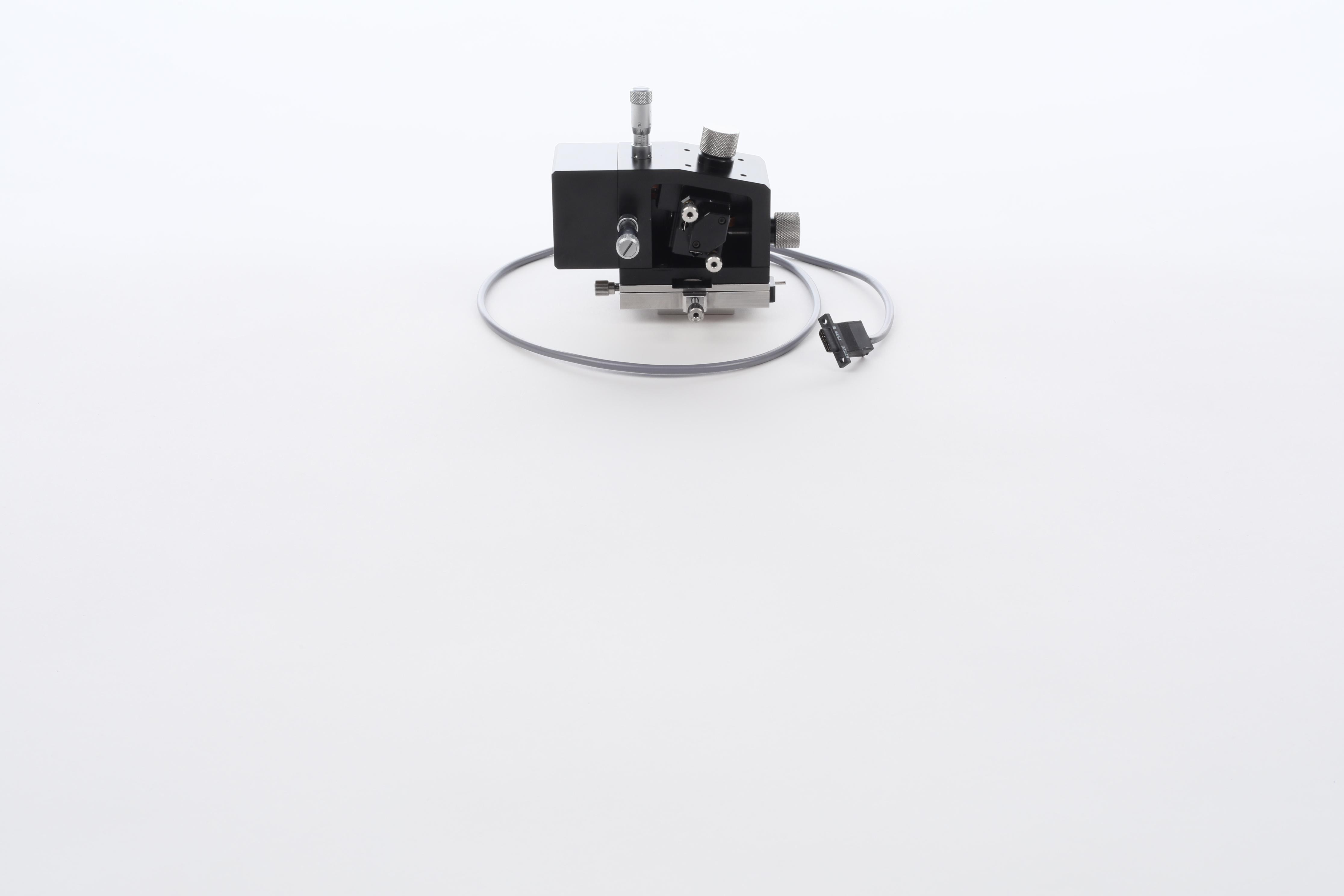
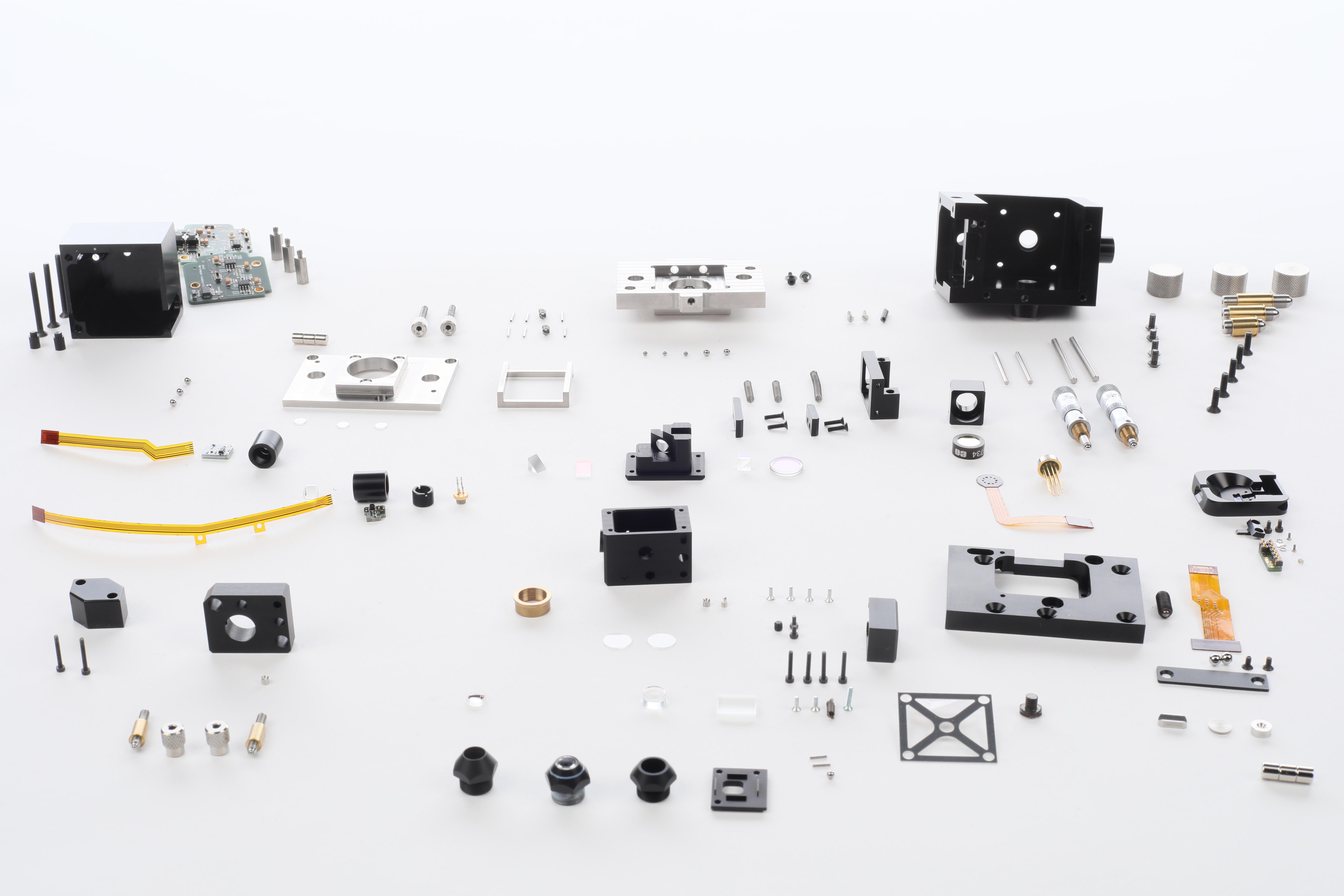
Researchers can come to EPFL to learn how to build the instrument from its spare parts. ©LBNI/EPFL
Using a new mode of atomic force microscopy, researchers at EPFL have found a way to see and measure protein assembly in real time and with unprecedented detail.
Proteins and molecules assemble and disassemble naturally as part of many essential biological processes. It is very difficult to observe these mechanisms, which are often complex and take place at the nanometer scale, far smaller than the normal visible range. At EPFL, however, an interdisciplinary team of researchers* has invented and applied a technique that allows these mechanisms to be examined with unprecedented precision. Their work is the subject of a paper published in Nature Nanotechnology.
Nanometric structures can only be seen with specialty microscopes, such as atomic force microscopes, which were invented in the mid-1980s. These instruments create an image by physically “feeling” the topography of the sample with an atomically sharp tip at the end of a tiny cantilever. The sample is then scanned point by point to create an image. As this takes time, only static samples can be imaged with conventional atomic force microscopes. However, this is of no use when scientists want to look at minute samples that change over time, such as protein assemblies.
“Change is essential for living matter and is therefore crucial to biological processes,” explains Prof. Georg Fantner, who leads EPFL’s Laboratory for Bio- and Nano- Instrumentation (LBNI). “So it was essential that we find a way to observe it.”

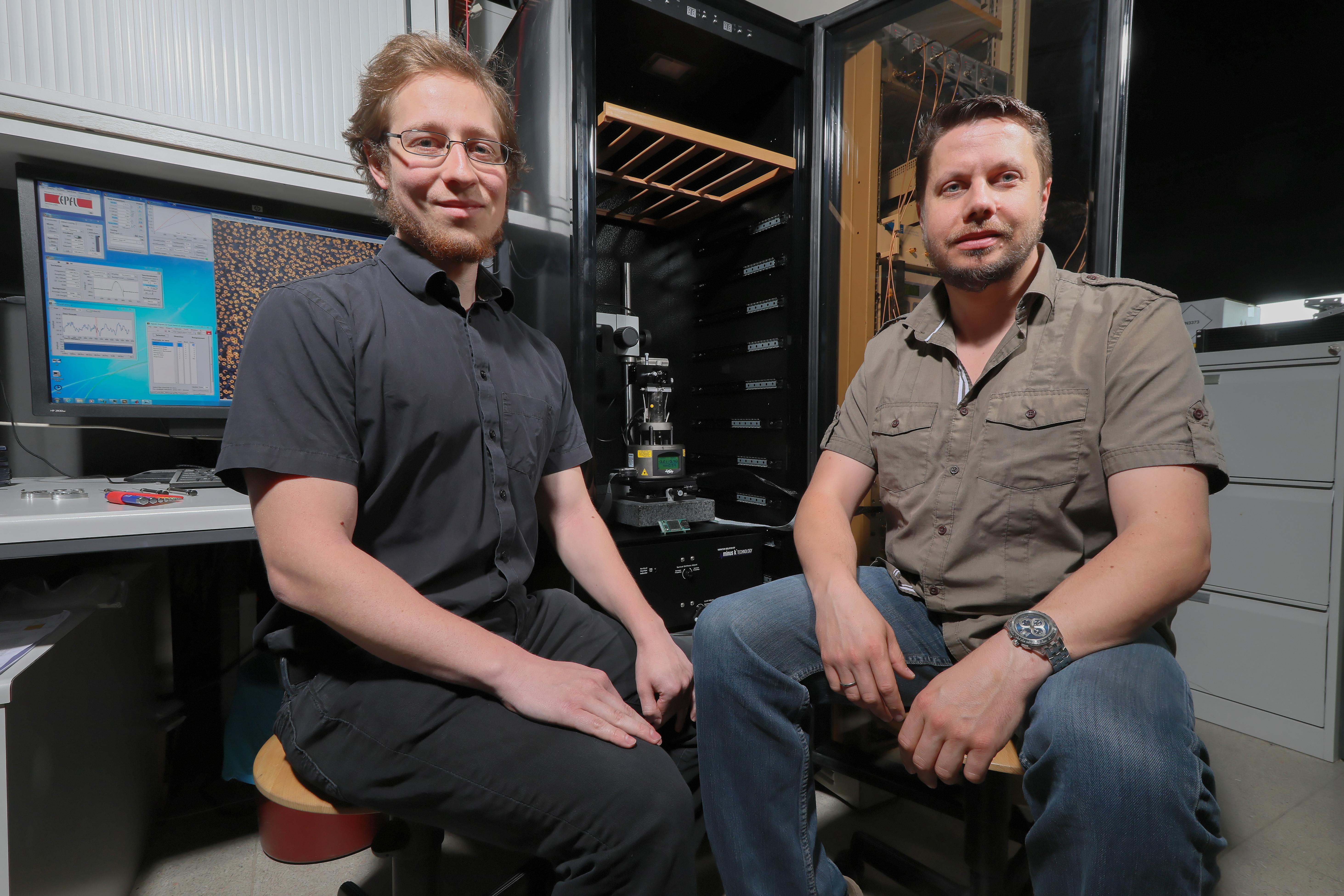
Adrian Nievergelt, doctoral assistant, and Georg Fantner, professor, with the microscope behind them.
To observe processes on a sample that changes over time, the scanning speed has to be increased. However, in traditional fast atomic microscopes, the forces exerted by the measurement can interfere with the molecular assembly process, especially since protein assemblies are often very fragile. The EPFL researchers found a method that solved the problem, by controlling the physical interaction of the sharp tip very precisely using pulsed laser light. This dramatically increased the scanning speed while preserving the gentle but extremely precise scanning motion.
2,000 lines per second
“We achieved this by using two lasers in the microscope, one of which is pointed at the base of the cantilever, locally heating it and thereby bending it slightly,” says Adrian Nievergelt, a PhD student at LBNI and co-first author of the paper. “By bending the cantilever, we can probe the surface much quicker, while still keeping fine control of the overall movement. In addition, we improved the overall system’s performance, allowing us to scan up to 2,000 lines per second.”
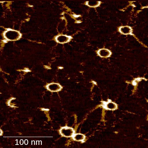
The researchers tested this new technology to analyze the dynamics of SAS-6 protein ring formation. This protein family plays a key role in the assembly of centrioles, which are minute organelles conserved from algae to men, fundamental for cell motility and division. As shown in the above video, the new instrument allowed the researchers for the first time to visualize the various stages of the ring assembly of SAS-6 proteins in real time. “This is a critical game changer for the field”, says Prof. Pierre Gönczy, an expert in centriole biology and co-author of the study. “Now we finally have a method to directly observe how this critical cellular component is assembled into a ring like polymer", adds Niccolò Banterle, a post-doctoral fellow in the Gönczy laboratory and co-first author of the study. "This allows us to better understand how nature controls the assembly of some of the smallest building blocks of life.”
Interested in this instrument? Come and make your own!
To share their new atomic force microscope (AFM) instrument in the spirit of open science, Georg Fantner, Adrian Nievergelt and co-workers decided to try something new. They designed a workshop for scientists from around the world who come to EPFL to learn how to build it.
Over 3 days, participants assemble their own device, and then test it to make sure it works fine. For the costs of a trip to EPFL and the microscope spare parts, they will leave with their own instrument. They can then start using it right away when they are back in their laboratory. The latest edition took place on campus in March with four participants from the UK, the USA and the Netherlands.
For Laia Pasquina Lemonche, a doctoral student at the University of Sheffield, having the opportunity to meet the inventors in person was of incredible value. “There is a real transfer of know-how. It’s amazing what we could do in 3 days. Even the best instruction manual cannot contain all the details we could get first-hand. Now I know exactly how it’s built, how it works and I will be able to fix it. I had never built anything before; I didn’t even know I loved building things!”
Luc Henry, scientific advisor.
* This research is the result of a collaboration between two EPFL entities: the Laboratory for Bio- and Nano- Instrumentation (LBNI) and Professor Pierre Gönczy’s Laboratory within the Swiss Institute for Experimental Cancer Research (ISREC) in the School of Life Sciences.
High-speed photothermal off-resonance atomic force microscopy reveals assembly routes of centriolar scaffold protein SAS‑6, Nature Nanotechnology, Adrian P. Nievergelt, Niccolò Banterle, Santiago H. Andany, Pierre Gönczy and Georg E. Fantner.
Images to download