Mini-colons revolutionize colorectal cancer research
In a breakthrough for cancer research, scientists at EPFL have created lab-grown mini-colons that can accurately mimic the development of colorectal tumors, offering a powerful new tool for studying and testing treatments for the disease.
As our battle against cancer rages on, the quest for more sophisticated and realistic models to study tumor development has never been more critical. Until now, research has relied on animal models and simplified cell culture methods, which are valuable but cannot fully capture the complex interplay of factors involved in tumor development.
Even newer, more advanced models for studying cancer, such as organoids – tiny, lab-grown versions of organs – do not faithfully replicate the cell behaviors and tissue architectures seen in actual tumors.
This gap has significantly hindered our understanding of the intricate processes underlying cancer initiation, progression, and response to treatment, and calls for more sophisticated models to accurately mimic the disease's complexity.
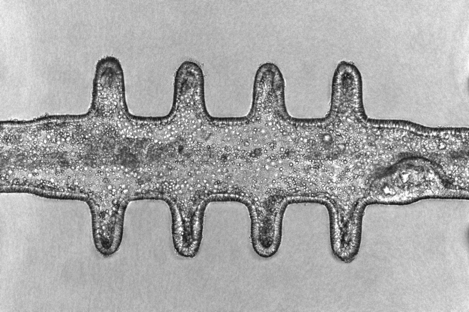
In a significant leap forward for cancer modeling, scientists have combined microfabrication and tissue engineering techniques to develop miniature colon tissues that can simulate the complex process of tumorigenesis outside the body with high fidelity, giving rise to tumors that closely resemble those found in vivo.
The breakthrough, now published in Nature, was made by Luis Francisco Lorenzo Martín, Tania Hübscher and other members of the group of Matthias Lütolf at EPFL, with input from the group of Freddy Radtke (EPFL) and colleagues at Roche’s Institute of Human Biology.
The mini-colons are topobiologically complex, meaning that they not only replicate the physical structure of colon tissue, including its distinctive crypt-and-lumen architecture, but they also mimic the cellular diversity present in the actual colon tissue during healthy and diseased states.
Optogenetics: Turning cancer “on”
Another important feature of the mini-colons is that they can be induced to develop tumors “at will” and in targeted areas – a massive advantage for cancer research. The researchers were able to turn inducible oncogenic genes on using “optogenetics”. This cutting-edge technique uses light to control biological processes such as gene expression.
By integrating a blue-light-responsive system into the mini-colons, the researchers made them undergo controlled oncogenic mutations, which can reveal tumor evolution with unprecedented details. This optogenetic approach allowed the scientists to induce targeted changes in specific cell populations within the mini-colons, mimicking the localized onset of colorectal cancer in the body.
“In essence, we used light to trigger tumorigenesis by turning on oncogenic driver mutations in a spatiotemporally controlled manner in healthy bioengineered colon epithelial organoids,” says Matthias Lütolf, who is also the founding director of Roche's new Institute of Human Biology. “This basically allows you to watch tumor formation in real-time and do very detailed analyses of a process that’s very difficult to study in a mouse.”
By manipulating genetic and environmental conditions, the researchers were also able to replicate and observe a range of tumor behaviors in the mini-colons, and even identified key factors influencing cancer progression – for example, the protein GPX2, which associated with stem cell characteristics and tumor growth.
This groundbreaking research offers a potent new tool for exploring the underlying mechanisms of colorectal cancer and testing potential therapies, particularly when applied to human patient-derived tissues. The mini-colons’ ability to mimic tumor dynamics can reduce our reliance on animal models, which can accelerate the discovery and development of effective treatments.
Other contributors
- EPFL Swiss Institute for Experimental Cancer Research (ISREC)
- Swiss Cancer Center Leman (SCCL)
- Institute of Human Biology, Roche Innovation Center Basel
Swiss Cancer League
Personalized Health and Related Technologies (PHRT)
EPFL
L. Francisco Lorenzo-Martín, Tania Hübscher, Amber D. Bowler, Nicolas Broguiere, Jakob Langer, Lucie Tillard, Mikhail Nikolaev, Freddy Radtke, Matthias P. Lutolf. Spatiotemporally resolved colorectal oncogenesis in mini-colons ex vivo. Nature 24 April 2024. DOI: 10.1038/s41586-024-07330-2
Images to download