Fundamentals of life: How centrosomes direct early embryos

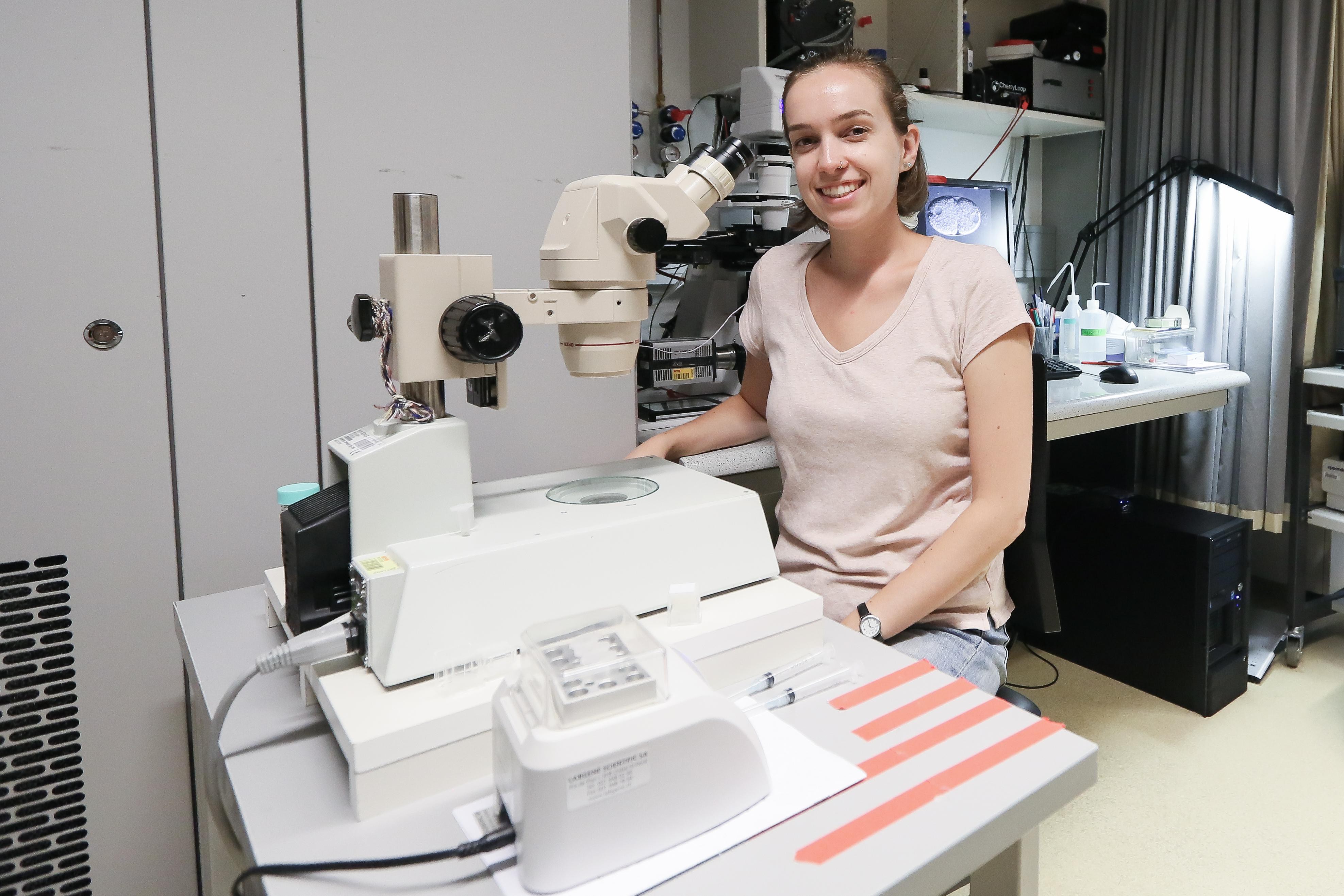
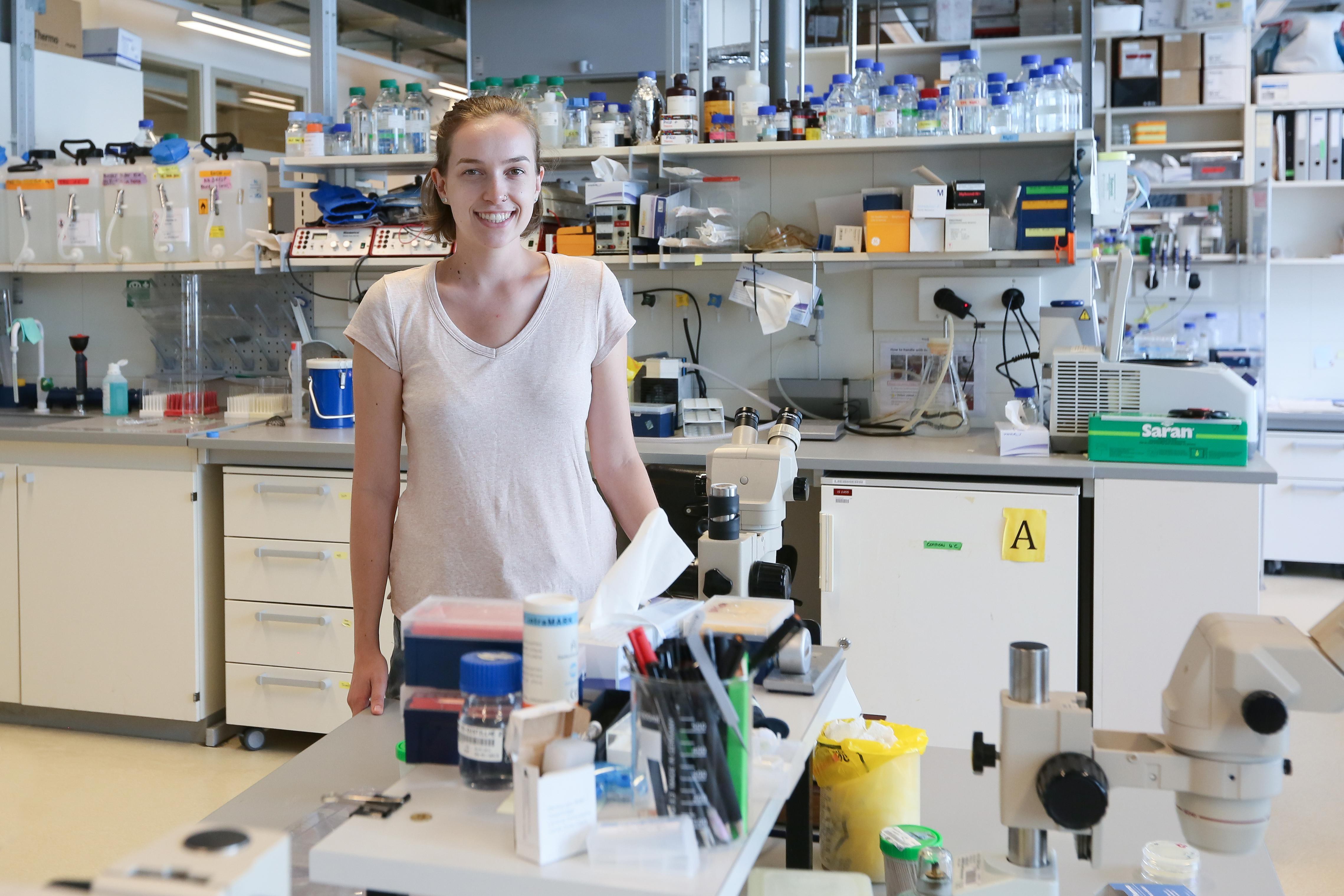
Sarah Herrman in the lab © Murielle Gerber/EPFL
Summer Series: Sarah Herrman is visiting Pierre Gönczy’s lab at EPFL from the University of California, Santa Cruz. Her project looks at how the cell’s centrosomes help organize the polarity and function of the developing embryo.
Centrosomes are cell organelles that are best known for their involvement in cell division. Each centrosome is made up of two small tubes arranged perpendicular to each other, called the centrioles. In turn, each centriole is made up of nine sets of microtubules.
When cell division begins, two centrosomes form at each pole of the cell and begin to assemble a spindle of fibers that will eventually split the cell’s genetic material (e.g. its chromosomes) in half, delivering a set to each of the two daughter cells.
But just as the North Pole differs from the South Pole, different regions of cells can have distinct features. This is called “cell polarity”, and it is essential in giving cells their unique identities. In developing embryos, for example, cell polarity ultimately dictates where the head of the organism will be located. This means that the cells of the early embryo divide asymmetrically, with certain proteins localized or produced unevenly inside them.
“For my project, we’re looking at cell polarization, which is the first thing that allows single-cell embryos of C. elegans to turn into complete, functioning organisms. There are data showing that the centrosome is somehow involved in setting up a polarity axis,” says Sarah Herrman, referring to the direction along which a cell will polarize.
To do this, she is using the roundworm C. elegans as a model organism – a popular tool among biologists. “C. elegans is a hermaphrodite, which means that it produces both sperm and eggs,” she says. “Right after fertilization, it produces a symmetric one-cell embryo with all of its cell components spread evenly throughout.” But to produce an organism with different types of cells —nerves, gut cells, a germ line — the embryo needs an asymmetric layout. This changes is known as “symmetry breaking,” and is the focus of both fundamental and medical research.
“We don’t know exactly what it is about the centrosome that makes it important for proper symmetry breaking, so that’s what my project is looking at,” says Herrman. More specifically, she is looking at the impact that centrosomes have on various proteins that play key roles in the polarization of many different cell types and organisms, including human beings. These proteins include a famous group of six “partitioning-defective” (PAR) proteins that are required for the first asymmetric cell division of the C. elegans zygote.
“What I am doing is knocking down the function of different proteins in C. elegans and analyzing how this affects the worm’s centrosomes – and therefore the polarity that they normally control.” Herrman is performing these knock-downs with either of two methods: C. elegans worms with a genetic mutation that prevents proper protein function, or RNA interference (RNAi), a Nobel-winning technique that targets mRNAs for degradation by exploiting the cell’s natural gene-controlling machinery.
“The mutants can be better because you really know that the gene is being knocked down,” says Herrman. But that doesn’t mean they are without limitations: “Some of the mutations are not compatible with adult life, so we can’t analyze their embryos.”
RNAi offers a way to work around these cases by knocking down the expression of specific genes during oogenesis and spermatogenesis. This allows Herrman to observe the effect on the resulting embryo’s centrosomes and its ability to polarize. “However, with RNAi you don’t necessarily have a complete knock down, so the two approaches are complementary.”
The worm embryos are also tagged with fluorescent proteins that allow observation of cell polarization. “We attach fluorescent labels to PAR proteins as well as other proteins involved in cell polarization, which allows us to visualize the centrioles and the posterior PAR domain,” says Herrman, referring to one area of the polarizing C. elegans embryo. “We also use tags that allow us to see the cortical contractions across the surface of the cell that move proteins toward the anterior end.”
So what’s in the future for Sarah Herrman? “I want to go to med school,” she says. “Maybe there’s a chance I’ll get an MD PhD, but I am more interested in the clinical side. Still, I think that research is something that all doctors should do, because if you don’t understand the research being done in the medical field, then you won’t be as effective in your practice.”
“I think it’s really important to do basic-biology research because it involves the fundamentals of all life,” she concludes. “It allows you to understand what’s going on normally in the cell and then from there you can develop applications more broadly.”
Sarah Herrman carried out her research as part of the Summer Research Program, an annual program for international undergraduate students run by EPFL's School of Life Sciences.
Images to download