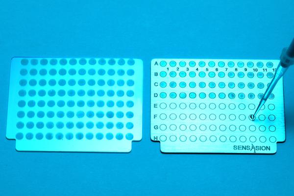
Electrochemistry flushes out antibiotic-resistant proteins

© 2018 EPFL Alain Herzog /Yingdi Zhu, Horst Pick
EPFL scientists, working in association with Valais Hospital in Sion and Fudan University in Shanghai, have developed a method for analyzing bacteria that – for the first time ever – lets doctors quickly see exactly which proteins are associated with antibiotic resistance.
Some bacteria contain proteins that indicate resistance to antibiotics. But to spot those proteins, doctors need to be able to open the bacteria membranes and analyze the proteins inside – something that until now has been impossible, since mass spectrometry can identify only small proteins. However, scientists at EPFL’s Sion campus working in association with colleagues at Fudan University in Shanghai have developed a single method that can be used to analyze a large spectrum of proteins and identify both the bacteria and their resistance to antibiotics. The process combines titanium dioxide nanoparticles with the energy emitted by UV light rays. Their research has been published in Chemical Science (an open-access journal of the Royal Society of Chemistry).
The WHO has named bacterial resistance to antibiotics as the single biggest threat to human health. It is the result of doctors excessively prescribing antibiotics, which has accelerated bacteria’s regular defense mechanisms, weeding out the weakest microbes and allowing the strongest to survive. Over time these bacteria have evolved to protect themselves against antibiotics by genetic mutation, passing genetic mutations on to their offspring or exchanging DNA with other bacteria.
Today scientists are trying to slow this process by better targeting specific bacteria to prevent new multi-resistant strains from forming. That means they must be able to precisely identify which bacteria they are dealing with. The main method used in hospitals to detect antibiotic resistance involves growing bacteria cultures in the presence of antibiotics, but this technique can take several hours or even days. Another method entails using mass spectrometry to analyze bacteria strains grown in a Petri dish; doctors place the bacteria on a steel plate and irradiate them with a laser. That generates a cloud of proteins that can be analyzed to determine the bacteria’s phenotype. But neither method is capable of identifying larger proteins – which is why the research team set out to develop a new one.
Steel plates imprinted with titanium dioxide
The EPFL scientists, working at the school’s Laboratory of Physical and Analytical Electrochemistry (LEPA), and their colleagues in Shanghai used steel plates that had been imprinted with titanium dioxide nanoparticles. “Titanium dioxide is a white powder that absorbs light. When hit with UV rays, the powder triggers an electrochemical reaction that enhances the laser’s effects by literally exploding open the bacteria membranes,” says Hubert Girault, head of LEPA.
This method opens up the bacteria much more than existing ones, releasing an array of biological molecules including protein, DNA, RNA, and lipids. “We looked mainly at proteins, since they are what can potentially alter or deteriorate the antibiotics,” says Horst Pick, a biologist who helped develop the method. “But we also found that we could use the same mass spectrometry technique to analyze all the other types of molecules released and obtain the bacteria’s ‘fingerprint.’ That can help doctors identify the specific type of bacteria.”
As a next step, the scientists hope to be able to work with the bacteria directly, without having to first grow cultures. That would shrink the analysis time to 30 minutes – a huge benefit since time is of the essence when fighting an infection – and ensure that doctors were targeting the right microbes. Highly promising trials have already been carried out.
Yingdi Zhu, Natalia Gasilova, Milica Jović, Liang Qiao, Baohong Liu, Lysiane Tissières Lovey, Horst Pick and Hubert H. Girault. Detection of antimicrobial resistance-associated proteins by titanium dioxide-facilitated intact bacteria mass spectrometry. http://pubs.rsc.org/en/Content/ArticleLanding/2018/SC/C7SC04089J