Cancer: banana peels can help identify the stages of melanoma

© 2016 Thinkstock
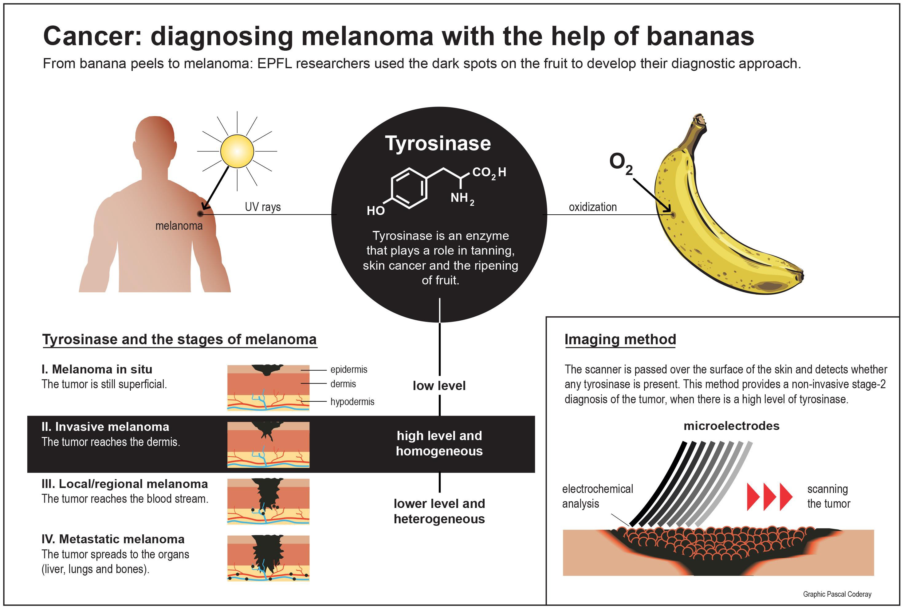
Human skin and banana peels have something in common: they produce the same enzyme when attacked. By studying fruit, researchers have come up with an accurate method for diagnosing the stages of this form of skin cancer.
When bananas age they become covered in black spots caused by the enzyme tyrosinase. It is a natural browning process common among certain organisms, including food. This same enzyme also plays a role in the type of skin cancer known as melanoma. This cancer’s tell-tale spots are the result of a malfunction in the regulation of tyrosinase. The malfunction disrupts the pigmentation of the skin by melanin, the body’s natural protection against the sun. Chemist Tzu-En Lin took advantage of the fact that tyrosinase occurs in both ripe fruit and human melanoma to develop an imaging technique capable of measuring tyrosinase levels and its distribution in human skin.
Research was first conducted on ripe fruit and then on samples of cancerous tissue. It was shown that the level and distribution of tyrosinase was indicative of the stage of the disease. It is not very apparent in stage 1; in stage 2 it is present in large amounts and evenly distributed; and in stage 3 it is unevenly distributed. The team, which is directed by Hubert Girault at the Laboratory of Physical and Analytical Electrochemistry at Sion (EPFL Valais Wallis), determined that this enzyme is a reliable marker of melanoma growth.
“The spots on human skin and on a banana peel are roughly the same size. By working with fruit, we were able to develop and test a diagnostic method before trying it on human biopsies,” said Dr. Girault.
The researchers made a scanner with eight microelectrodes lined up like the teeth of a comb and as flexible as the fingers of a hand. These tiny sensors pass over the uneven surface of the skin without damaging it and measure the electrochemical response within an area of a few square millimeters. The electrodes calculate the quantity and distribution of tyrosinase, which allows the researchers to determine the stage of the melanoma. This system could obviate the need for invasive tests like biopsies.
The next step will be to use this same scanner to view the tumors and eliminate them. “Our initial laboratory tests showed us that our device could be used to destroy the cells,” said Dr. Girault.
_ _ _
This work was carried out by a research group at EPFL Valais Wallis, an EPFL satellite campus based in the canton of Valais.
The results of the study were published in Angewandte Chemie.
Tzu-En Lin, Alexandra Bodarenko, Andreas Lesch, Horst Pick, Fernando Cortés- Salazar, Hubert H. Girault.