At the Dubochet Center for imaging, atoms are made visible
Fully operational for two weeks, the Dubochet Center for imaging was presented to the press on November 22. It promises considerable advances in biomedical research thanks to the precision of the images that its microscopes can obtain.
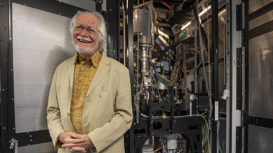
“Imagine how an old man might feel when a Center like this is named after him...” The emotion could be heard, yesterday, in the voice of Jacques Dubochet. The 2017 Nobel Prize in Chemistry was participating in the official launch of the Dubochet Center for Imaging (DCi), a joint platform of EPFL, the University of Lausanne and the University of Geneva.
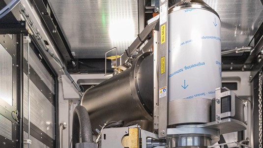
The Center has more than enough to put the Lake Geneva region on the world map of advanced imaging facilities. “We have here the best model of microscope that money can buy. And we have not one, but three,” said Henning Stahlberg, director of the DCi.
These impressive machines, priced in the millions of francs, make use of the discovery that earned Dubochet and his colleagues the prestigious award: cryo-electron microscopy, or cryo-EM for short. “We have developed a technique by which the samples to be studied are instantly vitrified, instead of being frozen,” said Dubochet. This speed avoids the formation of ice crystals, which would be harmful to the molecules to be observed. “The properties of water in this state are still mysterious,” he added. Once stabilized in this way, the samples can withstand the flow of electrons that pass through them and are “photographed” without being altered.
One center, two sites
The DCi has five of the most powerful electron microscopes in the world and will be available to all researchers in the region. In addition, there is a prototype dedicated to the development of new technologies that will further improve image quality.
The most powerful microscopes are temporarily based between the UNIL and EPFL campuses, in the Cubotron building. Two other machines are installed in Geneva. “There are many synergies between the two sites and permanent collaborations,” explains Robbie Loewith, Deputy Director of the DCi and head of the Geneva office. “The pooling of all these resources allows us to obtain results with unparalleled speed and precision.”
Understanding molecular interactions
As a clear example, the DCi has published the complete structure of a ferritin molecule – responsible for the transport of iron in the blood – with a precision of 1.4 Å: a degree of detail that makes it possible to observe each atom individually and its connections with neighboring atoms. The same is true for the SARS-CoV-2 Spike protein, which can now be “walked around” atom by atom.
“This is extremely valuable for biomedical research,” says Henning Stahlberg. Because we can understand how biological proteins interact with each other, we can also develop molecules that can block or promote these interactions – for example, in the case of cancer treatments using chemotherapy.
In other words, thanks to this new center, cryo-electron microscopy is now reaching a degree of maturity that will allow considerable scientific advances and for which Western Switzerland will be at the forefront.