Protein elasticity underlies Alzheimer's and Parkinson's disease
")
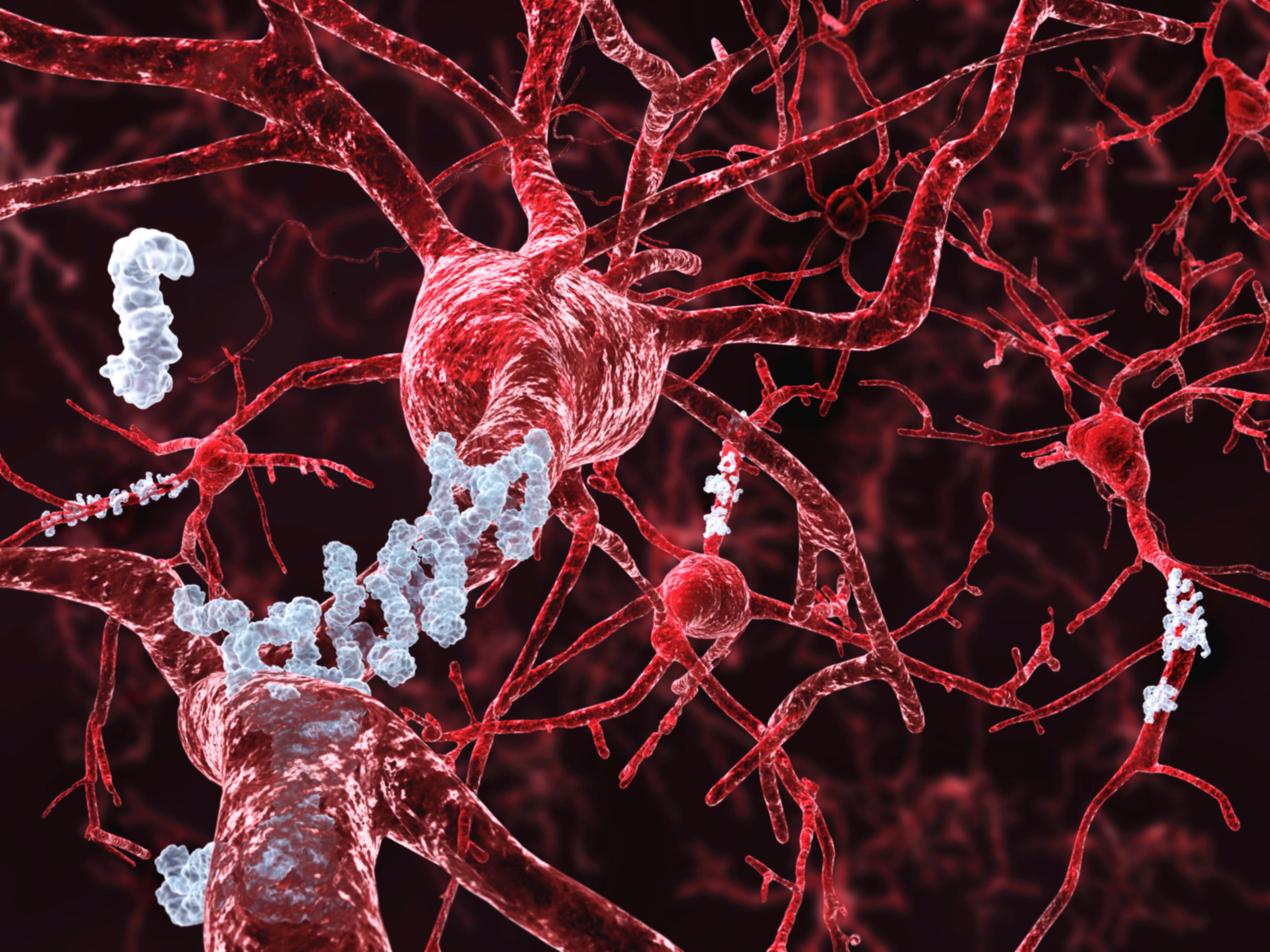
Amlyoid beta aggregating outside neurons. (Image: ThinkStock)
Researchers from EPFL have studied the evolution of amyloid-beta and alpha-synuclein from peptides to fibrillar aggregates, and have mapped out how gradually the fibril’s stiffness increases in the process.
Alzheimer’s disease is characterized by the presence of extracellular amyloid plaques in the brain, which are formed when the small amyloid-beta peptide aggregates into fibrils, which then cluster together to form plaques. A similar aggregation of the protein alpha-synuclein is linked to Parkinson’s disease. Consequently, research for treating these diseases focuses heavily on understanding amyloid fibril and plaque formation. Now, researchers at EPFL have mapped out how amyloid-beta and alpha-synuclein aggregates gradually stiffen as they evolve from peptides to fibrils. Their findings, which are published in Angewandte Chemie, could lead to new pharmacological strategies to fight Alzheimer’s and Parkinson’s, and might even contribute to areas of food science and biotechnology that utilize other types of amyloid fibrils.
In neurodegenerative disorders like Alzheimer’s and Parkinson’s disease, proteins with normal physiological functions begin to mis-fold and clump together into insoluble aggregates. A research group led by Giovanni Dietler at EPFL investigated the mechanical changes of this process by combining conventional methods for studying protein aggregates – fluorescence imaging and circular dichroism – with atomic force microscopy. Here, the microscope consists of a cantilever with a sharp tip that scans the surface of specimens, producing high-resolution images and “stiffness maps” down to the nanometer scale.
Using this approach, the scientists scanned samples of amyloid-beta and alpha-synuclein aggregates prepared by the lab of Hilal Lashuel at EPFL’s Brain & Mind Institute. The samples represented the various stages of evolution from single peptide to oligomer and fibril. As the cantilever on atomic force microscope measured the force that its scanning tip applied on the sample, the scientists were able to detect how the mechanical properties of the samples changed. Specifically, they were able to measure how they became increasingly stiff as they aggregated into more complex forms. This change in elasticity is described by what is known as “Young’s modulus”.
The increasing stiffness of the protein aggregates reflects an internal change that takes place in the 3D structure of amyloid-beta and alpha-synuclein. When they are single peptides and proteins respectively, they both have a coiled structure. But as they begin to aggregate, they adopt a complex internal structure known as beta-sheet, which is a mesh of anti-parallel peptides, and seems to be a key factor in the increasing rigidity of amyloid plaques.
The findings provide a novel, mechanical view on protein fibrils that contributes to elucidate their stability, toxicity, and also potential for clearance in treating associated disorders. Taking into consideration changes in elasticity could lead to new therapeutic approaches in treating neurodegenerative diseases like Alzheimer’s and Parkinson’s.
This study represents a collaboration between EPFL’s Laboratory of Physics of Living Matter and Laboratory of Molecular & Chemical Biology of Neurodegeneration, with ETH Zürich Department of Health Sciences and Technology.
Reference
Ruggeri FS, Adamcik J, Jeong JS, Lashuel HA, Mezzenga R, Dietler G. Influence of the β-Sheet Content on the Mechanical Properties of Aggregates during Amyloid Fibrillization.Angewandte Chemie 14 January 2015. DOI: 10.1002/anie.201409050