Glass slides that stand to revolutionize fluorescence microscopy

© 2020 EPFL / Alain Herzog
EPFL scientists have developed a new type of microscope slide that can boost the amount of light in fluorescence microscopy by a factor of up to 25. These new slides can both amplify and direct light, making them ideal for applications ranging from early-stage diagnosis to the rapid archiving of pathology samples.
For scientists, the glass slides used to prepare samples for looking at under a microscope are part and parcel of their work – and they haven’t changed much in nearly 200 years.
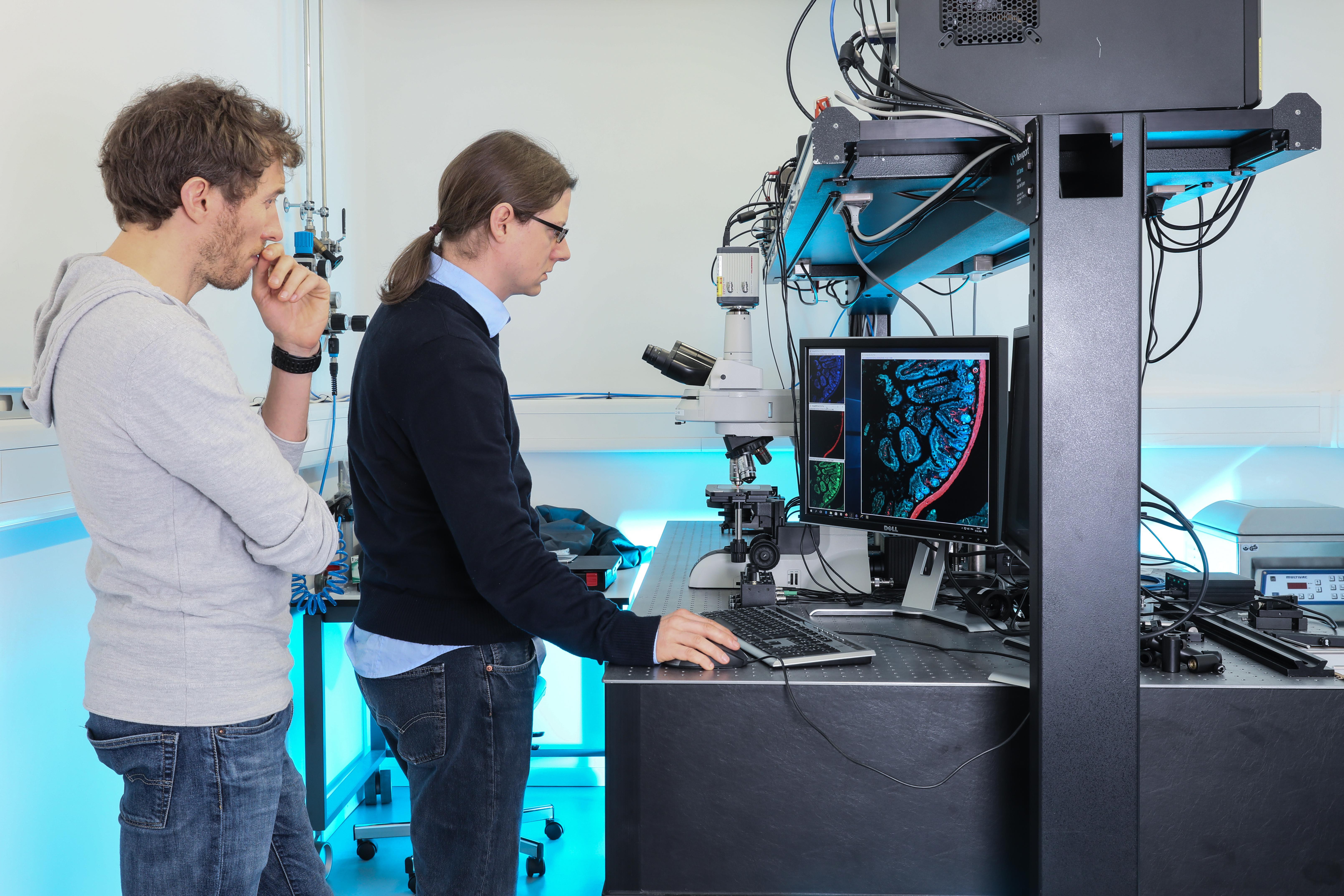
At the Institute of Microengineering in Neuchâtel, part of EPFL’s School of Engineering, researchers have developed a novel type of glass slide that stands to break with tradition. Their slides have a coating that “structures” light, allowing for up to 25 times more light to be emitted and thereby enhancing the sensitivity of the microscopes they are used with.
Nicolas Descharmes and Raphaël Barbey developed their slides specifically for fluorescence microscopy, which is widely used to diagnose cancer and autoimmune diseases, identify allergies or sequence DNA. Their design has unique optical properties and it allows the detection of minute amount of light. This could be especially useful for making early-stage diagnosis, quickly identifying specific types of cancer and rapidly archiving pathology samples. “In an ideal case, our slides could be used to detect the presence of one molecule, where 25 molecules would be needed on conventional slides” says Descharmes.
The scientists have patented their technology, and their slides – which have already been used by researchers in a range of fields – will soon be tested at several companies. The pair has received the support from EPFL, the Gebert Rüf foundation and Innosuisse, and plans to launch their own company in the coming months. Through their startup, Descharmes and Barbey will be able to scale up production and make the slides available to hospital labs and diagnostics providers.
Eliminating two main drawbacks
Fluorescence microscopy works by detecting the light that compounds called fluorophores emit when excited. More specifically, fluorophores absorb light at a given wavelength, called the excitation wavelength, and, in response, emit light at a longer wavelength, called the emission wavelength. With fluorescence microscopes, scientists can view objects that are naturally fluorescent or that have been marked with a fluorophore, and that would be impossible to see with a regular microscope.
But there are two main drawbacks to using glass slides in fluorescence microscopy. First, fluorophores usually emit a very small amount of light. And second, most of the light that they do emit gets lost in the slide, meaning it can’t be used. As a result, many compounds are hard or even impossible to detect unless there is a fairly large amount in the sample.
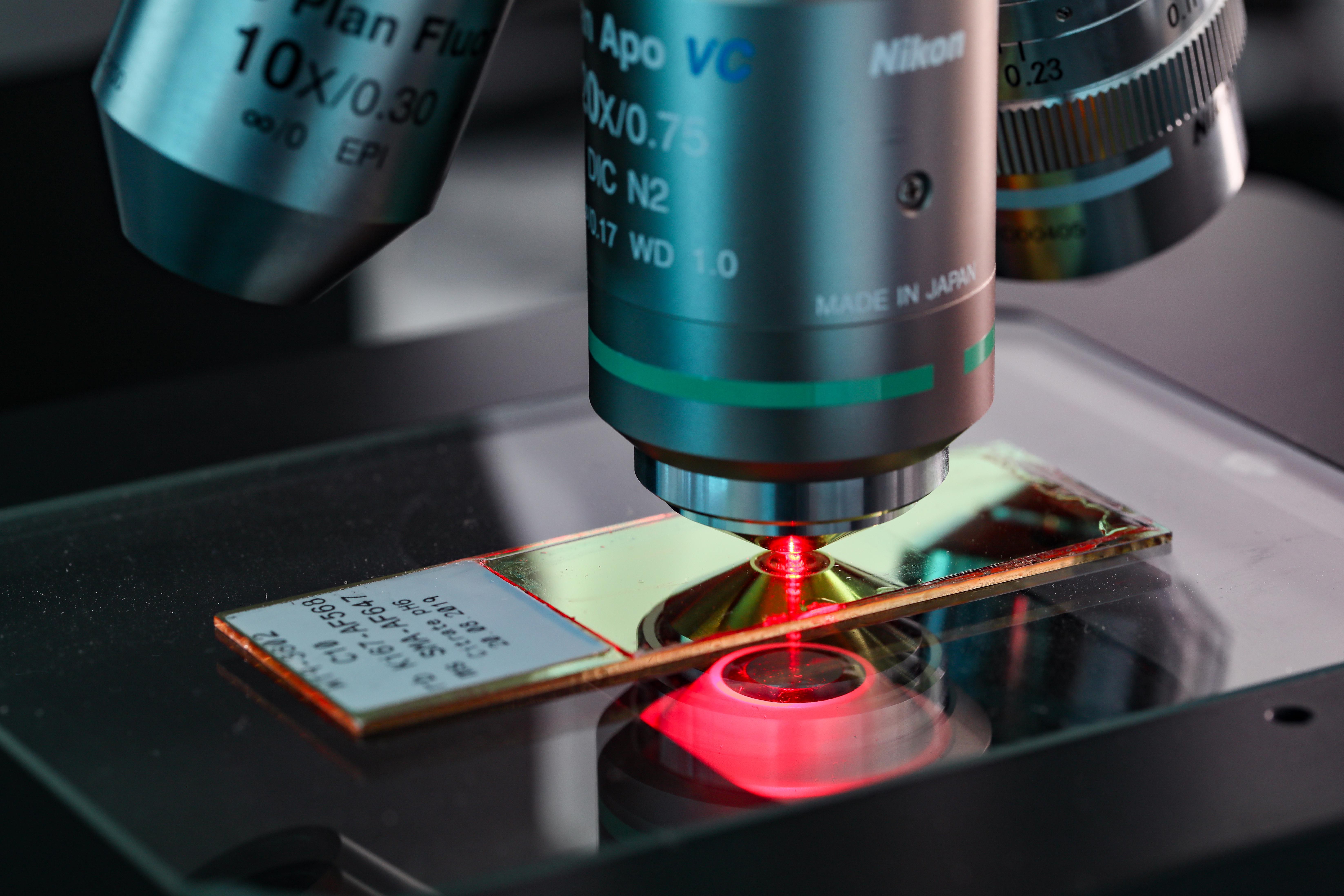
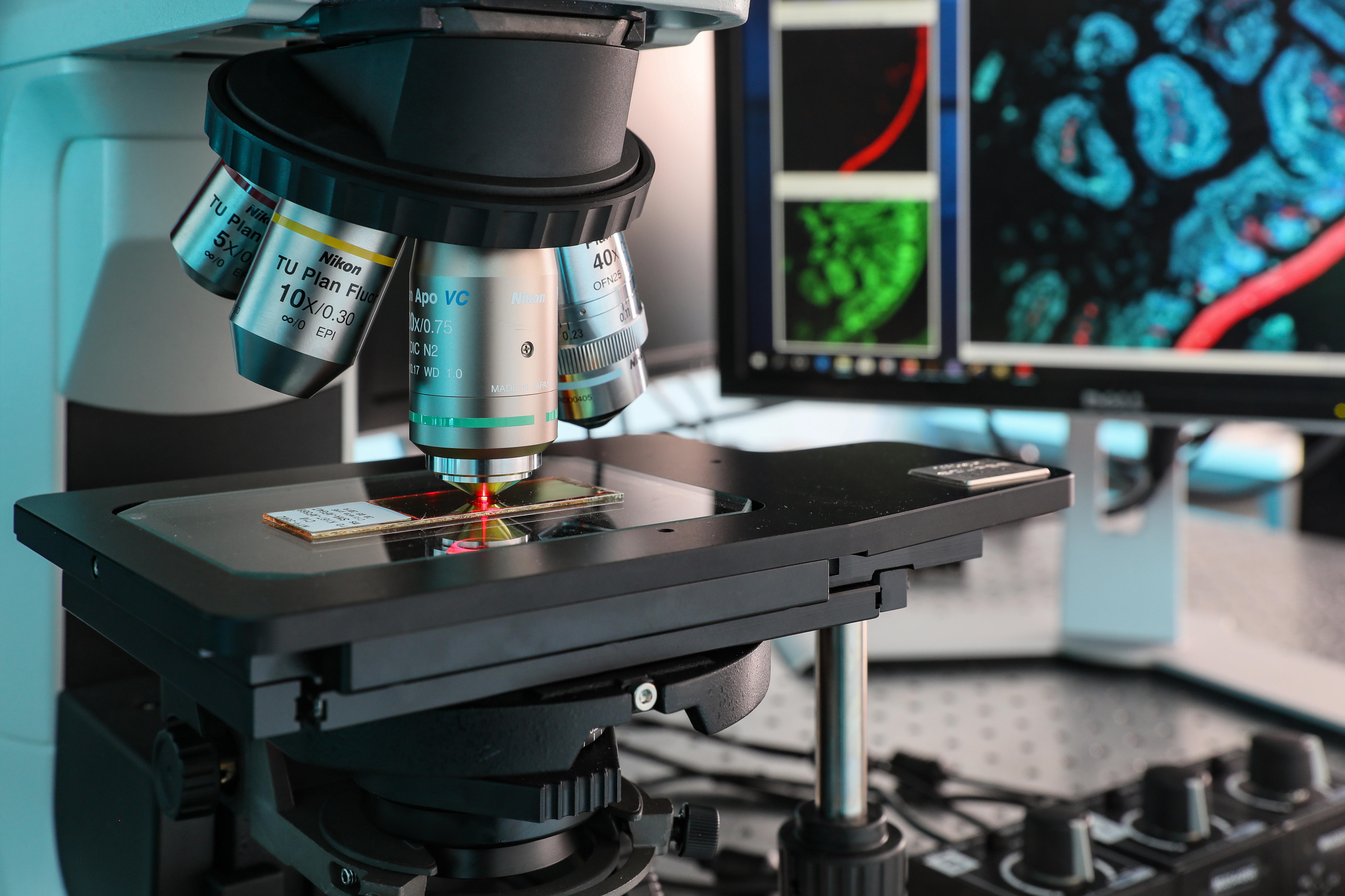
An optical layer cake
Descharmes and Barbey’s slides have a layered structure that is capable of controlling the electromagnetic environment surrounding the samples. When light is shined on the fluorophores in a sample, they emit more light than they would on a conventional slide, and all of that light is directed towards the microscope’s detector. That results in images that are clearer or that can be generated more quickly.
“What I’ve seen so far is highly promising,” says Séverine Lorrain, a senior technician at UNIL’s Protein Analysis Facility who works on detecting proteins in samples. “I was really impressed by how efficient the slides are in amplifying the fluorescence signal. That means I could avoid going through a separate signal amplification step – a big advantage since that step often introduces background noise.”
Jessica Dessimoz, head of the EPFL Histology Core Facility, also finds the new slides promising: "The surface of these slides improves the visualization of the fluorescent signal and reduces the exposure time required. It could prove very useful for applications like cyclic immunofluorescence. "
Enabling early-stage diagnosis
The EPFL scientists are targeting several applications for their invention, such as the early-stage diagnosis of some types of cancer or the easier reading and archiving of histopathology slides, which are commonly used in the analyses of biopsies. According to Barbey, “Scanning conventional slides in fluorescence takes a long time because the signals are weak. But with our slides, the process could go a lot faster. The hard part will be convincing researchers to give up some of their old slides!” Raphaël Barbey is currently working on the industrialization of the production of these slides with another Neuchâtel flagship of technology – the CSEM (Swiss Center for Electronics and Microtechnology).
These new slides mark a shift in the field of fluorescence microscopy. Nearly all microscope parts have been continuously optimized in recent decades, except for the slides. The light sources are now more powerful, the cameras are more sensitive and the lenses of better quality. “Surprisingly, the slides have been a little bit forgotten in this improvement process,” says Barbey. “The advantage of our approach is that it entails a minor change for microscope users but could substantially improve their instruments’ performance.”
Nicolas Descharmes currently works at the Photovoltaics and thin film electronics laboratory of EPFL (Neuchâtel) and Raphael Barbey is now a CSEM employee.
Images to download