Unraveling the light of fireflies

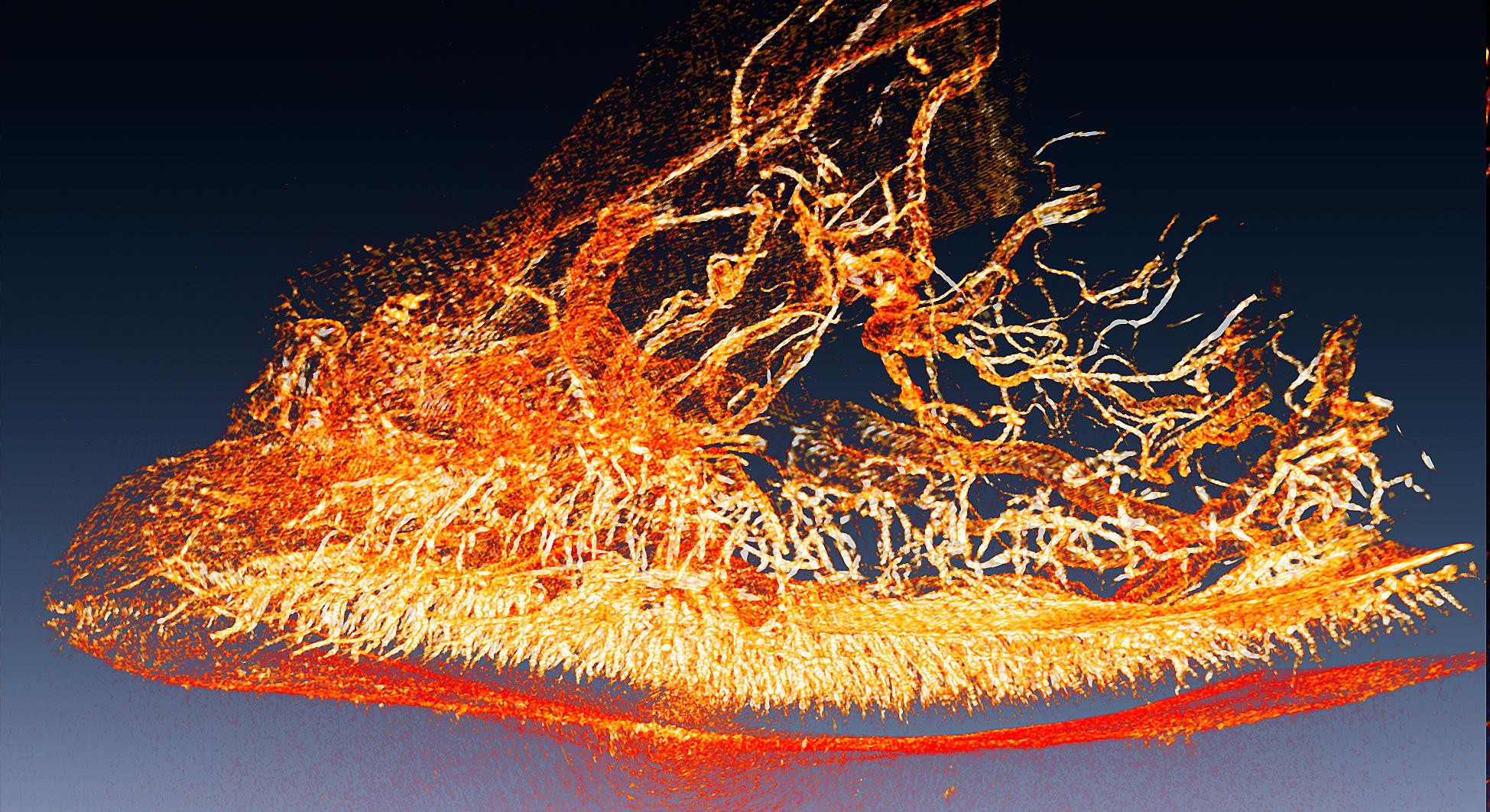
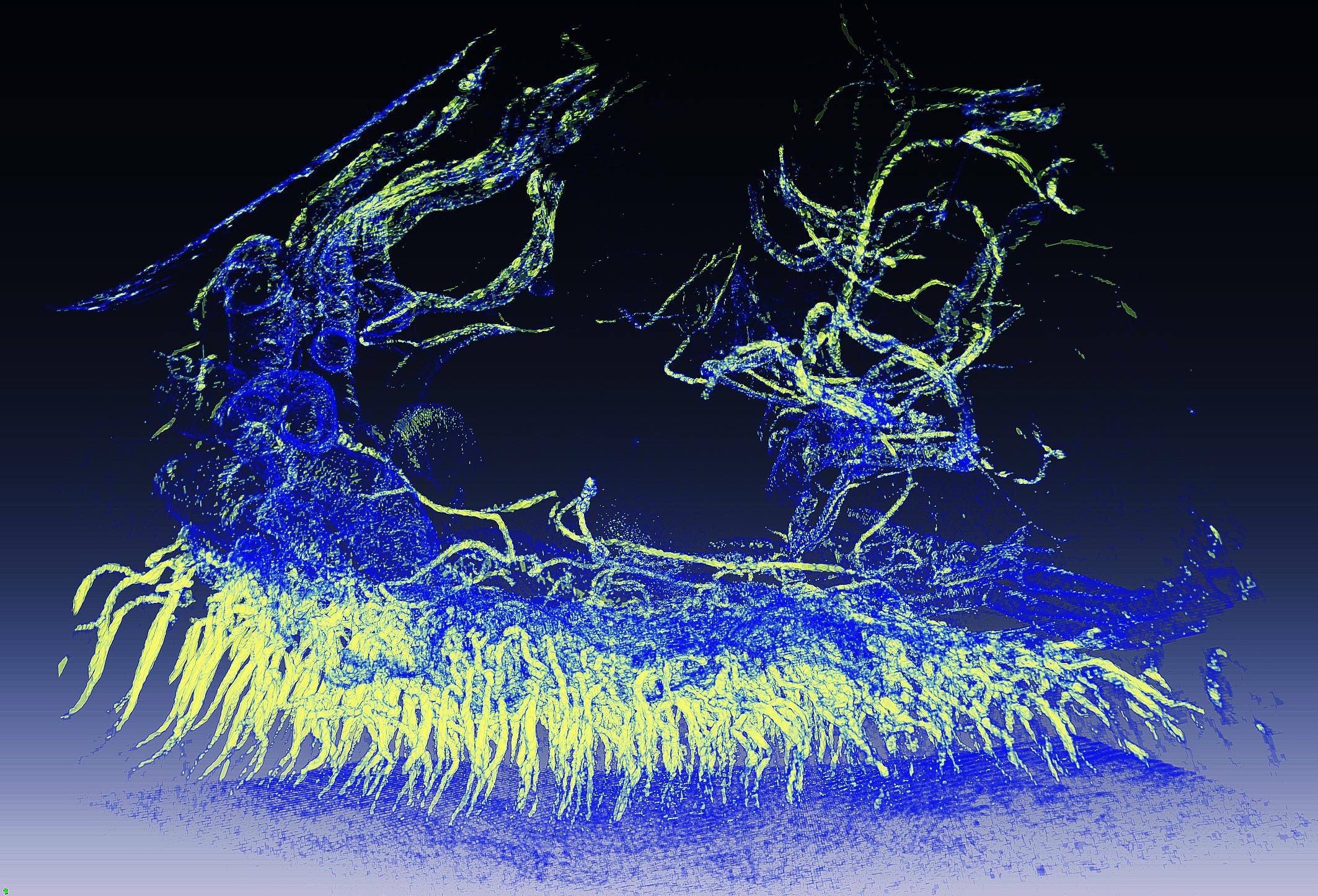
The firefly's lantern organ ©2014 G. Margaritondo/EPFL
How do fireflies produce those mesmerizing light flashes? Using cutting- edge imaging techniques, scientists from Switzerland and Taiwan have unraveled the firefly’s intricate light-producing system for the first time.
The firefly’s light-producing organ is called the “lantern”, and it is located in the insect’s abdomen. It looks like a series of tubes progressing into smaller ones and so one, like a tree’s branches growing into twigs. The function of these tubes, called, is to supply oxygen to the cells of the lantern, which contain luciferase and can produce light. However, the complexity of the firefly’s lantern has made it difficult to study this mechanism in depth, and reproduce it for technological applications.
Giorgio Margaritondo at EPFL, Yeukuang Hwu at the Academia Sinica and their colleagues at the National Tsing Hua University in Taiwan have successfully used two sophisticated imaging techniques to overcome the complexity of the firefly lantern and map out how oxygen is supplied to light-emitting cells. The techniques are called synchrotron phase contrast microtomography and transmission x-ray microscopy. They can scan down to the level of a single cell, even allowing researchers to look inside it.
By applying these techniques on live fireflies, the scientists were able to see the entire structure of the lantern for the first time, and to also make quantitative evaluations of oxygen distribution.
The imaging showed that the firefly diverts oxygen from other cellular functions and puts it into the reaction that breaks up luciferin. Specifically, the researchers found that oxygen consumption in the cell decreased, slowing down energy production. At the same time, oxygen supply switched to light emission.
The study is the first to ever show the firefly’s lantern in such detail, while also providing clear evidence that it is optimized for light emission thanks to the state-of-the-art techniques used by the scientists. But Margaritondo points out another innovation: “The techniques we used have an advantage over, say, conventional x-ray techniques, which cannot easily distinguish between soft tissues. By using an approach based on changes in light intensity (phase- contrast) as opposed to light absorption (x-rays), we were able to achieve high- resolution imaging of the delicate firefly lantern.”
This work represents a collaboration of EPFL with the following institutes in Taiwan: Academia Sinica, the National Tsing Hua University, the Endemic Species Research Institute, the National Taiwan University, and the National Cheng Kung University.
Reference
Tsai Y-L, Li C-W, Hong T-M, Ho J-Z, Yang E-C, Wu W-Y, Margaritondo G, Hsu S-T, Ong EBL, Hwu Y. Firefly Light Flashing: Oxygen Supply Mechanism. Phys. Rev. Lett. 113, 258103 17 December 2014. DOI: 10.1103/PhysRevLett.113.258103
Images to download