How a waste product of exercise protects neurons from trauma damage

ThinkStock photos
Researchers led by EPFL have found how lactate, a waste product of glucose metabolism can protect neurons from damage following acute trauma such as stroke or spinal cord injury.
Stroke or spinal cord injury can cause nerve cells to receive excessive stimulation, which ultimately damages and even kills them. This process is known as excitotoxicity, and it is one of the reasons why time following such trauma is critical, while it is also implicated in progressive neurodegenerative diseases, e.g. Alzheimer’s disease. A team of scientists led by EPFL has now discovered that lactate, which is produced in the brain and even muscles after intense exercise, can be used to protect neurons against excitotoxicity. The study is published in the Nature journal Scientific Reports.
Following acute trauma such as a stroke or spinal cord injury, a certain type of receptors go into overdrive and overwhelm the target neuron with a barrage of electrical signals. This causes a build-up of calcium ions inside the neuron, which triggers toxic biochemical pathways that ultimately damage or kill it.
The receptors that cause this are called NMDA receptors, and interact with the neurotransmitter glutamate. NMDA receptors are a major target in research and medicine, as they are implicated in a number of disorders, including epilepsy, schizophrenia, Parkinson’s and even Alzheimer’s.
A team of researchers led by Pierre Magistretti from EPFL and the King Abdullah University of Science and Technology, investigated the effects of glutamate on cultured neurons from the brains of mice. The scientists used a new, non-invasive imaging technique called Digital Holographic Microscopy that can visualize cells structure and dynamics with nanometer-level resolution.
Previous studies have suggested that lactate could protect neurons against excitotoxicity. Lactate is produced in the brain and in muscles after intense exercise as a waste product of glucose metabolism. Nonetheless, how lactate protects neurons has eluded scientists until now.
The researchers tested the effects of glutamate on the mouse neurons with and without lactate. The results were revealing: glutamate killed 65% of the neurons, but when with lactate, that number dropped to 32%.
The researchers then aimed to determine how lactate protects neurons. By using different receptor blockers on the mouse neurons, they determined that lactate triggers the production of ATP, the cell’s energy molecule. In turn, the produced ATP binds and activates another type of receptor in the neuron, which turns on a complex cascade of defense mechanisms. As a result, the neuron can withstand the onslaught of signals from the NMDA receptor.
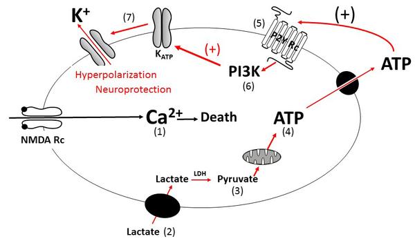
(1) Excessive glutamate activity triggers a strong influx of calcium (Ca2+) into the neuron through NMDA receptors, which leads to cell death. (2) Lactate is transported into the neuron and (3) converted to pyruvate by the enzyme lactate dehydrogenase (LDH). (4) Pyruvate is then transported into mitochondria by the mitochondrial pyruvate carrier (MPC) where it generates ATP. (5) ATP is then released through pannexins and activates the receptor P2Y, which (6) activates the PI3K pathway. (7) This triggers the opening of potassium channels (K+), which causes the neuron to hyperpolarize, decreasing the neuron's excitability, and thus protecting it from excitotoxic damage.
The breakthrough can advance our understanding of neuroprotection, which could lead to improved pharmacological ways to ameliorate the irreparable damage caused by stroke, spinal cord injury, and other trauma.
---
This work involved a collaboration of EPFL’s Brain Mind Institute with the King Abdullah University of Science and Technology, and the University Hospital of Lausanne (CHUV). It was funded by the FNRS and the NCCR Synapsy.
Reference
Jourdain P, Allaman I, Rothenfusser K, Fiumelli H., Marquet P, Magistretti PJ. L-Lactate protects neurons against excitotoxicity: implication of an ATP-mediated signaling cascade.Scientific Reports 6:21250, 19 February 2016. DOI: 10.1038/srep2125019